
A Man with torn biceps does preacher curls. Working out and injury rehabilitation after 6 months of an untreated grade 3 full bicep tear.

Depict the salivary glands and ducts within the oral cavity using X-ray illustration.
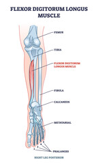
Flexor digitorum longus muscle with human leg and foot bones outline diagram. Labeled educational physiology scheme with phalanges and metatarsal skeletal and muscular system vector illustration.
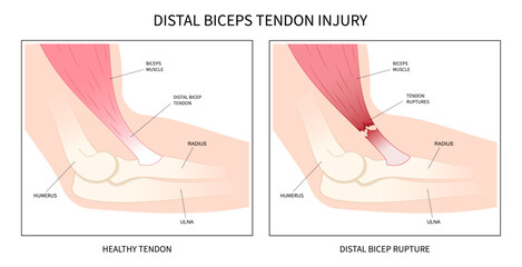
distal bicep tendonitis rotator cuff pain upper arm inflamed
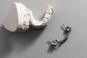
Dental casting gypsum model of human jaws. Crooked teeth and distal bite. Shots were made before treatment with braces . Technical shots on gray background.
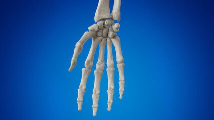
3d rendered illustration of the hand bones
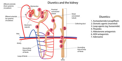
Diuretics and the kidney. Vector illustration

Film X ray wrist radiograph show distal forearm bone broken ( distal end radius fracture). The patient has wrist pain, swelling and deformity. Medical imaging for investigation and technology concept
3D imaging of the human hand bones and joints, hightech medical detail, clipart isolated on a white background
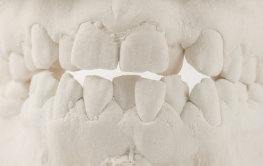
Dental casting gypsum model of human jaws. Crooked teeth and distal bite. Shots were made before treatment with braces . Technical shots on gray background
Human adult hand bones x-ray image. Medical and anatomy radiography or imagery

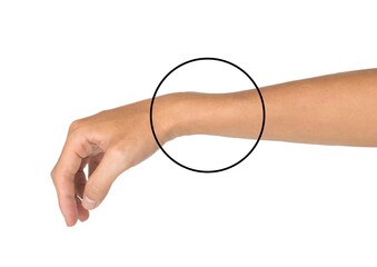
man on the beach with a bandage in his wrist

A 3D model of the human skeleton, highlighting the spinal column for orthopedic educational purposes
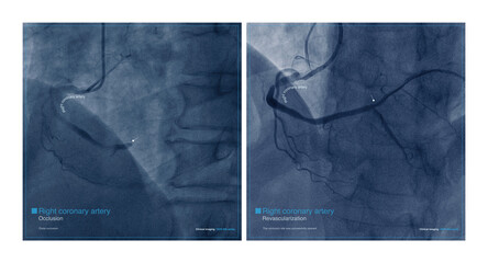
Male, 68 years old, chest pain for 7 hours. Coronary angiography suggests occlusion of the distal right coronary artery. The patient was successfully placed with a coronary stent.
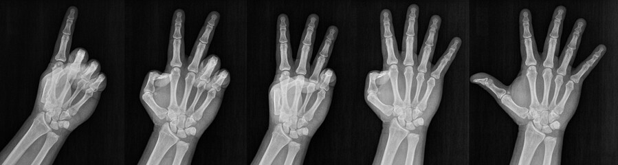
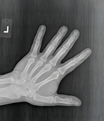
Film xray x-ray or radiograph of a hand and fingers showing the numbers one through five 1-5. One, two, three, four five in gestural language, manual communication, or signing aka sign language
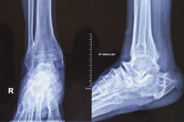
X-ray of ankle joint both view. Diffuse sclerosis at tarsal, metatarsal bones and distal shaft of tibia and fibula with deformed shaped. joint spaces are markedly reduced.
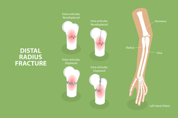
3D Isometric Flat Vector Conceptual Illustration of Distal Radius Fracture, Labeled Educational Diagram
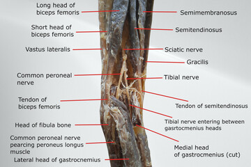
anatomy of popliteal region with the distal portion of back of thigh and proximal portion of back of leg. picture containing related muscles, nerves, tendon and fossa.
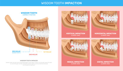
Wisdom Tooth Impaction Infographics
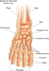
Medical illustration of the main parts of the bones of the foot in anterior view, with annotations.
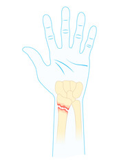
Colles Fracture is a complete fracture of the radius bone close to wrist. Broken wrist.
A photo of plain radiograph on dark background in hospital. The film use for diagnosis the illness of patient.Medical concept. A children with fracture distal ulnar bone. A green stick fracture.
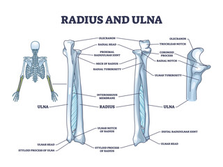
Radius and ulna bone anatomy with arm skeletal structure outline diagram. Labeled educational scheme with upper body parts and hand long bones vector illustration. Detailed physiological description.
Avascular necrosis hand displacement break painful radial dislocated fall onto an outstretched closed fragment of dorsal Ulna Extra Intra articular and De quervain's proximal by car pole anatomy

Goldilocks Buttercup (Ranunculus auricomus). Cauline Leaf Closeup
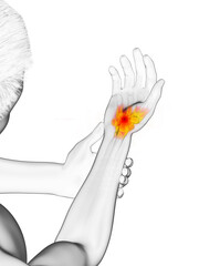
3d rendered medically accurate illustration of a man having a painful wrist

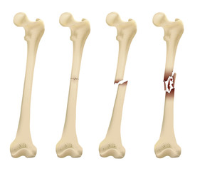
Structure and components of long bone. Proximal epiphysis, Distal epiphysis, Diaphysis. Compact bone and an inner medullary cavity containing bone marrow.

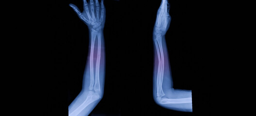
Film X-ray wrist radiograph show lower end of forearm bone broken (distal end radius fracture) from traffic accident. Highlight on broken site and painful area. Medical imaging and technology concept
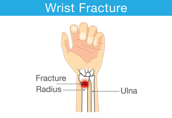
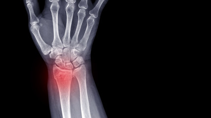
Diagram of wrist have bone fracture

Drawind of human brain on chalkboard with inscription distal
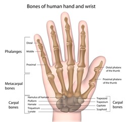
Bones of the hand, labeled.
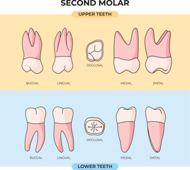
collection of upper and lower Second Molar tooth anatomy in various angles
3D illustration of the human foot, detailing bones, joints, and muscles, used in podiatry and sports medicine, 3D Illustration
Opthalmoscope or Otoscope icons set in flat style
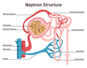
Nephron structure. Urine formation organ, functional unit of the kidney.
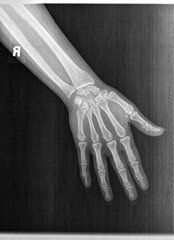
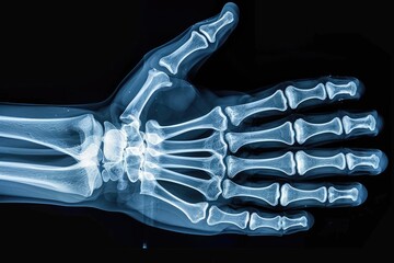
Film xray or radiograph of a normal left hand of an adult male. AP view show human's hand. normal bone structure of all phalanges carpal bones metacarpal, distal radius and ulna. joint space is normal

Illustration of human foot with ankle pain. anatomy, physical therapy concept
Human adult female right hand bones x-ray image. Medical and anatomy radiography or imagery
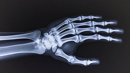
Human adult hand bones x-ray image. Medical and anatomy radiography or imagery

Keep nails healthy by tending to cuticles and hangnails

Gastroduodenoscopy distal end and manipulator in the hands of doctors in treatment room
Femur Bone element vector illustration
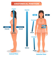
Anatomical positions vector illustration. Scheme of superior, inferior and proximal, distal locations, as well as medial, lateral and anterior, posterior sides
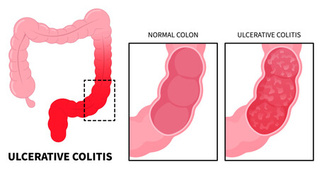
Anatomy of large intestines inflammation with ulcerative colitis and Crohn's disease that has ulcer painful or diarrhea
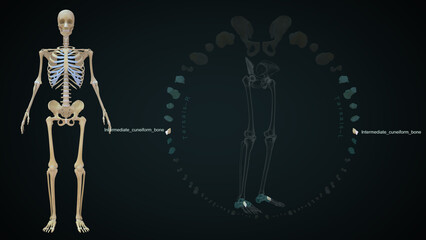
The medial cuneiform is one of the cuneiforms, it is the most medial in the distal row of tarsal bones 3d illustration

School age girl renting an electric scooter or bicycle using smartphone, making contactless payment through mobile app. Bicycle rental in the city in the autumn-winter season
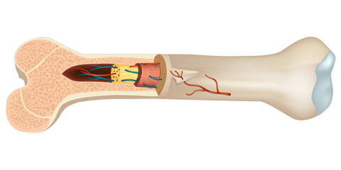
Structure and components of long bone. Proximal epiphysis, Distal epiphysis, Diaphysis. Compact bone and an inner medullary cavity containing bone marrow.
Film X-ray wrist radiograph show lower end of forearm bone broken (distal end radius fracture) from falling. Highlight on broken site and painful area. Medical imaging and technology concept
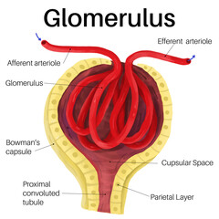
The glomerulus is a network of small blood vessels.
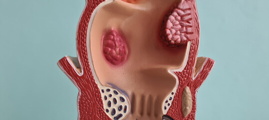
Anatomical model of rectum with hemorrhoids closeup
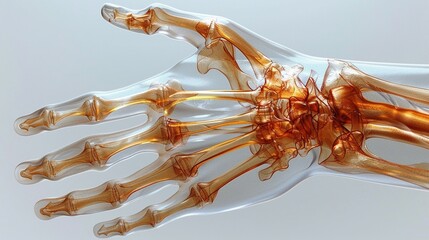
3D imaging of the human hand bones and joints, hightech medical detail, clipart isolated on a white background
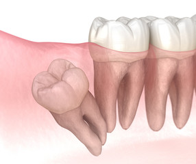
Distal impaction of Wisdom tooth. Medically accurate tooth 3D illustration
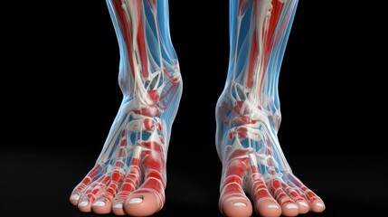
3D illustration of the human foot, detailing bones, joints, and muscles, used in podiatry and sports medicine
Opthalmoscope or Otoscope icons set in monochrome
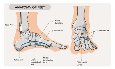
Arches of the feet. Foot skeleton anatomy.
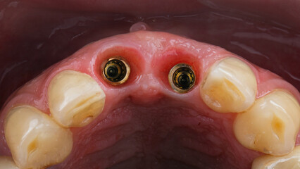
excellent gingival cavity with two dental implants, view through the distal mirror

Illustration of human foot with ankle pain. anatomy, physical therapy concept
Film xray or radiograph of a normal left hand of an adult male. AP view show human's hand. normal bone structure of all phalanges carpal bones metacarpal, distal radius and ulna. joint space is normal
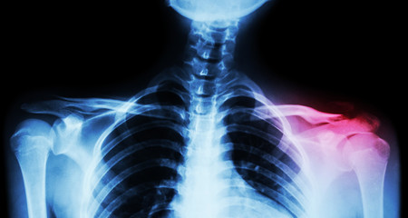
Film x-ray both clavicle AP ( front view ) : show fracture distal left clavicle
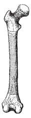
Femur, The shaded part represents the diaphysis, vintage illustration.
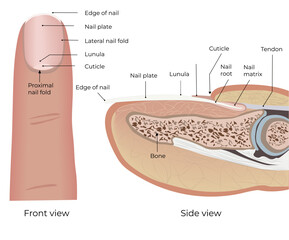
Structure of nail and finger. Medical illustration. Anatomical illustration. Suitable for doctors, students, medical journals, articles. Material for podiatrists. Nail Health. Educational material for

Male, 68 years old, chest pain for 7 hours. Coronary angiography suggests occlusion of the distal right coronary artery. The patient was successfully placed with a coronary stent.
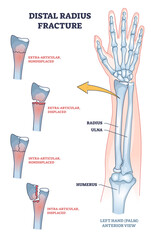
Distal radius fracture and broken arm bone types anatomy outline diagram. Labeled educational scheme with extra articular nondisplaced and displaced radius and ulna comparison vector illustration.